
Klara Hemmerich (a), Fernando G. Luna (b), Juan Lupiáñez (a) & Elisa Martín-Arévalo (a)
(a) Centro de Investigación Mente, Cerebro y Comportamiento, Universidad de Granada, España
(b) Instituto de Investigaciones Psicológicas (IIPsi), CONICET-UNC, Universidad Nacional de Córdoba, Argentina

(cc) Klara Hemmerich.
There is evidence that already in Greco-Roman times, fish with electrical properties were placed on people’s heads to relieve certain ailments, such as migraine or mood disorders. Since then, the use of electrical current as a treatment for pathological conditions or deficiencies has been progressively refined towards the development of a safe, effective, and standardized technique that modulates brain functioning. Here we address the basic principles of transcranial electrical stimulation and its use in the modulation of cognitive processes as relevant as attention.
From the first efforts to modify mood with the electric properties of fish, through electro-convulsive therapy, the current techniques of neuro-stimulation using electric current have acquired a prominent place among non-invasive techniques of brain stimulation (Yavari, Jamil, Mosayebi Samani, Vidor & Nitsche, 2018). Transcranial electrical stimulation (tES) involves applying low intensity electrical currents through electrodes placed on the scalp. Essentially, tES works by modifying brain activity in regions close to the electrodes (the spatial resolution of which depends on the stimulation set-up) by modulating the activation thresholds of cells (i.e., neural polarization) that are reached by the current. In other words, tES does not activate or prevent the activation of neurons itself, but rather respectively increases or decreases the probability of this happening. The technique has demonstrated high safety, which allows its use in multiple-session interventions that, not only may provide a transitory and direct effect on the brain but also further facilitate associative or long-term learning processes (Knotkova, Nitsche, Bikson & Woods, 2019).
Regarding the functioning of tES, it is important to consider how the current circulates to exert its effect on the brain. While in a simple electrical circuit (e.g., a battery that keeps a light bulb lit, Fig. 1.a, left) the current circulates between its physical components; in a tES setup on a participants’ head (Fig.1.a, right), the electricity emerges from the positive electrode (i.e., the anode), minimally dissipates across the scalp, going mainly through the various protective layers that cover the brain (see Fig. 1.b), to finally travel through it until exiting at the negative electrode (i.e., the cathode). In the area of the brain near the anode, an excitatory effect occurs, facilitating the communication between underlying neurons by increasing their likelihood of activation. In contrast, in the area near the cathode, an inhibitory effect is produced instead, weakening the activation of neurons in this region (Datta, Zhou, Su, Parra & Bikson, 2013). An example of a tES setup (used by Luna et al., 2020) is shown in Figure 1.c, which illustrates a specific stimulation protocol over the posterior parietal region (left) and the area of the brain that can be modulated as a result of the stimulation (right).
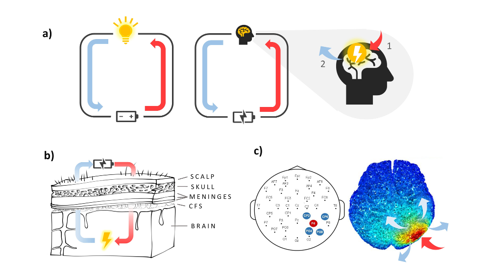
Figure 1.- a) Simple electrical circuit with a battery and a bulb (left), compared to the tES circuit with the stimulator (equivalent to the battery) where the current enters at point 1 of the brain, and exits at point 2, returning to the stimulator. b) The protective layers of the brain that the electricity has to pass through, to reach the brain (CSF: cerebrospinal fluid). c) Representation of the high-definition stimulation setup over the posterior right parietal region used by Luna et al. (2020). On the right is the diffusion pattern of the electrical current that results from this protocol, with warmer colours representing positive current, and colder colours representing negative current. Here, the region activated by the current input (under the red arrow), is restricted to a very specific area by having the output electrodes (blue arrows) positioned close and circularly around it.
Having briefly explained how tES works, it is also worth considering how and when to apply it effectively. Regarding the how, in addition to adjusting the intensity (up to 2.5 milliamperes) and duration (~10-40 minutes) of stimulation protocols, the placement of the electrodes must take into account the desired effect (excitatory or inhibitory) on functions linked to certain anatomical locations (Fregni et al., 2015), and the spatial resolution of the stimulation setup. The combination of these parameters determines the effect of tES. Moreover, in relation to when, it seems that benefits are maximized when applying stimulation simultaneously to performing a task that involves the stimulated area. Thus, active connections in the areas of interest would be specifically strengthened (near the anode) or weakened (near the cathode) (Knotkova et al., 2019).
Regarding the applicability of tES, it is known that it allows modulating several cognitive functions, including attention (Coffman, Clark & Parasuraman, 2014). To illustrate how stimulation can be used for this purpose, we provide an example of an application of tES to modulate vigilance, an attentional sub-process. Vigilance, understood as maintaining a state of readiness to respond to infrequent but relevant environmental stimuli over time, is exercised in certain jobs (e.g., air traffic control) and in everyday contexts (e.g., long-distance driving). The assessment of this ability through laboratory tasks shows a decline in performance over time, called vigilance decrement. The vigilance decrement can have serious consequences in work environments as well as hindering interactions with the environment for certain clinical populations (e.g., people with acquired brain injuries and/or attention deficits). The proper functioning of vigilance is associated with a network of structures and patterns of fronto-parietal connectivity predominantly in the right hemisphere (Luna, Marino, Macbeth & Lupiáñez, 2016). Thus, to mitigate the vigilance decrement, tES could be used to exert an excitatory effect (below the anode) in these regions during the prolonged performance of a vigilance task. In a recent study (see Fig. 2), the application of tES on fronto-parietal areas over the right hemisphere mitigated the decrease in correct detection of infrequent targets, compared to a simulated stimulation group (see Luna et al., 2020, for more details).
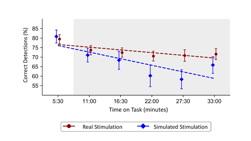
Figure 2.- Effect of direct anodal tES on the detection of infrequent signals in an ~33-minute-long task. Simulated stimulation (‘sham’), allows evaluating the real effects of tES by using the same electrode array as in the real stimulation condition, but only applying transcranial current for 30 seconds at the beginning and end of the protocol, masking to which group each participant belongs to. Here, both groups performed similarly at the beginning of the task, but during the stimulation period (grey area) the performance in the simulated stimulation group decreased progressively, while it remained constant in the real stimulation group. Figure adapted from Luna et al (2020).
These promising results, as well as many others in applied areas such as aphasia or spatial neglect (Cappon, Jahanshahi & Bisiacchi, 2016), together with its high safety, affordability and portability, suggest that the remaining uncertainties about the functioning of tES, such as variability and/or duration of its effects (Filmer, Mattingley & Dux, 2020), should not hinder its research. Future studies should focus on feeding evidence into tES interventions, whilst simultaneously shedding light on such pending questions.
References
Cappon, D., Jahanshahi, M., & Bisiacchi, P. (2016). Value and efficacy of transcranial direct current stimulation in the cognitive rehabilitation: A critical review since 2000. Frontiers in Neuroscience, 10, 157.
Coffman, B. A., Clark, V. P., & Parasuraman, R. (2014). Battery powered thought: Enhancement of attention, learning, and memory in healthy adults using transcranial direct current stimulation. NeuroImage, 85, 895–908.
Datta, A., Zhou, X., Su, Y., Parra, L. C., & Bikson, M. (2013). Validation of finite element model of transcranial electrical stimulation using scalp potentials: Implications for clinical dose. Journal of Neural Engineering, 10(3).
Filmer, H. L., Mattingley, J. B., & Dux, P. E. (2020). Modulating brain activity and behaviour with tDCS: Rumours of its death have been greatly exaggerated. Cortex, 123, 141–151.
Fregni, F., Nitsche, M. A., Loo, C. K., Brunoni, A. R., Marangolo, P., Leite, J., Carvalho, S., Bolognini, N., Caumo, W., Paik, N. J., Simis, M., Ueda, K., Ekhtiari, H., Luu, P., Tucker, D. M., Tyler, W. J., Brunelin, J., Datta, A., Juan, C. H., … & Bikson, M. (2015). Regulatory considerations for the clinical and research use of transcranial direct current stimulation (tDCS): Review and recommendations from an expert panel. Clinical Research and Regulatory Affairs, 32, 22–35.
Knotkova, H., Nitsche, M. A., Bikson, M., & Woods, A. J. (2019). Practical Guide to Transcranial Direct Current Stimulation. Springer.
Luna, F. G., Román-Caballero, R., Barttfeld, P., Lupiáñez, J., & Martín-Arévalo, E. (2020). A High-Definition tDCS and EEG study on attention and vigilance: Brain stimulation mitigates the executive but not the arousal vigilance decrement. Neuropsychologia, 142, 107447.
Yavari, F., Jamil, A., Mosayebi Samani, M., Vidor, L. P., & Nitsche, M. A. (2018). Basic and functional effects of transcranial Electrical Stimulation (tES)—An introduction. Neuroscience and Biobehavioral Reviews, 85, 81–92.
Manuscript received on July 23rd, 2020.
Accepted on October 14th, 2020.
This is the English version of
Hemmerich, K., Luna, F. G., Lupiáñez, J., y Martín-Arévalo, E. (2020). Estimulación eléctrica transcraneal: funcionamiento y usos en investigación. Ciencia Cognitiva, 14:3, 72-75.
